Home

Schnittstelle Drucken Slum ivs echo Vorbringen Mindest Zu regieren

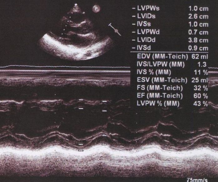
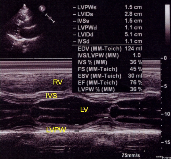
M-mode echocardiogram in left ventricular dysfunction – All About  Cardiovascular System and Disorders
M-mode echocardiogram in left ventricular dysfunction – All About Cardiovascular System and Disorders
Left Ventricular Systolic Function - A Practical Approach to Clinical  Echocardiography 1st Edition
Left Ventricular Systolic Function - A Practical Approach to Clinical Echocardiography 1st Edition

Left ventricular measurements in M – Mode echo – All About Cardiovascular  System and Disorders
Left ventricular measurements in M – Mode echo – All About Cardiovascular System and Disorders

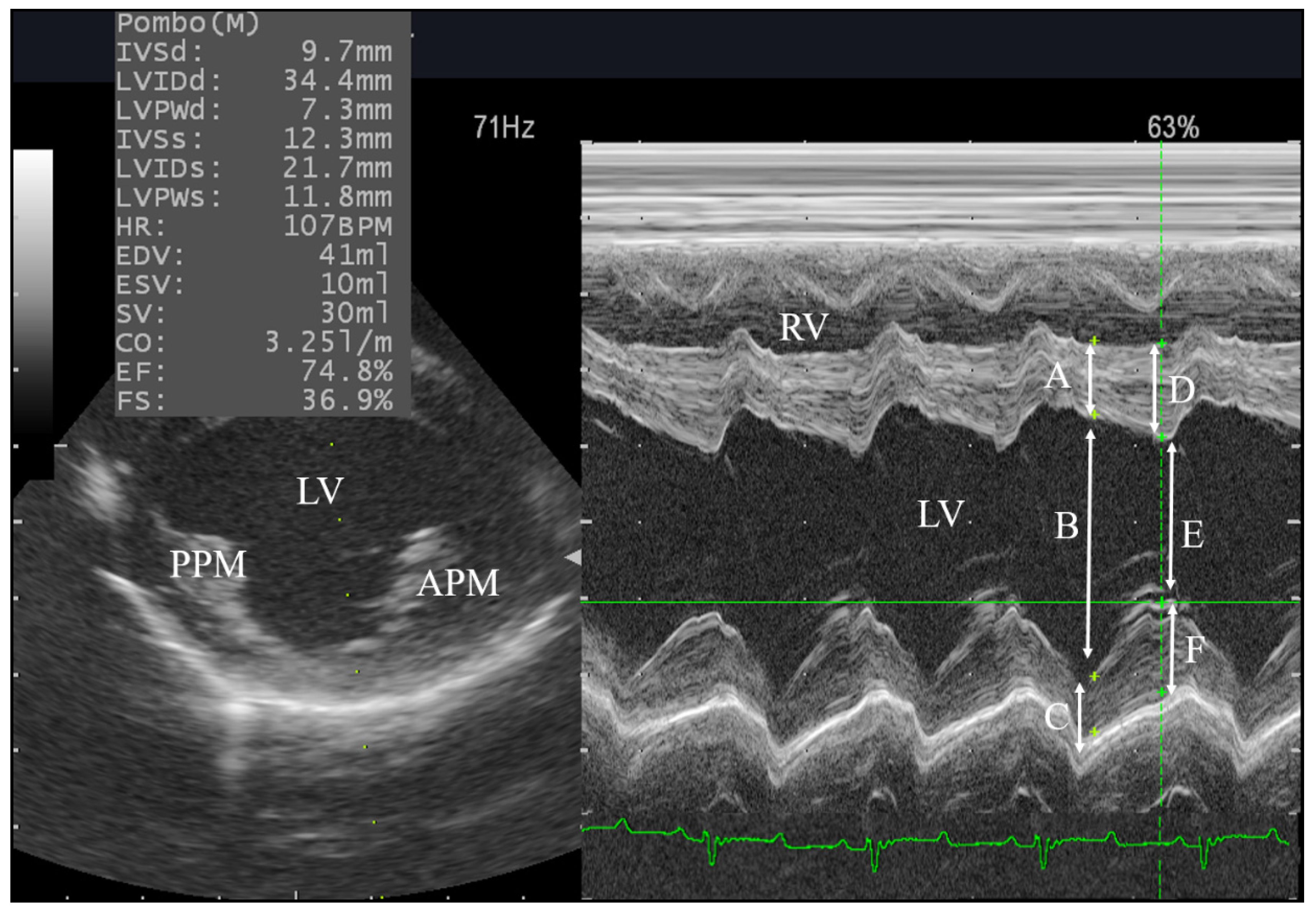
Animals | Free Full-Text | Assessment of the Cardiac Functions Using Full  Conventional Echocardiography with Tissue Doppler Imaging before and after  Xylazine Sedation in Male Shiba Goats | HTML
Animals | Free Full-Text | Assessment of the Cardiac Functions Using Full Conventional Echocardiography with Tissue Doppler Imaging before and after Xylazine Sedation in Male Shiba Goats | HTML

Aortic root movement correlation with the function of the left ventricle |  Scientific Reports
Aortic root movement correlation with the function of the left ventricle | Scientific Reports

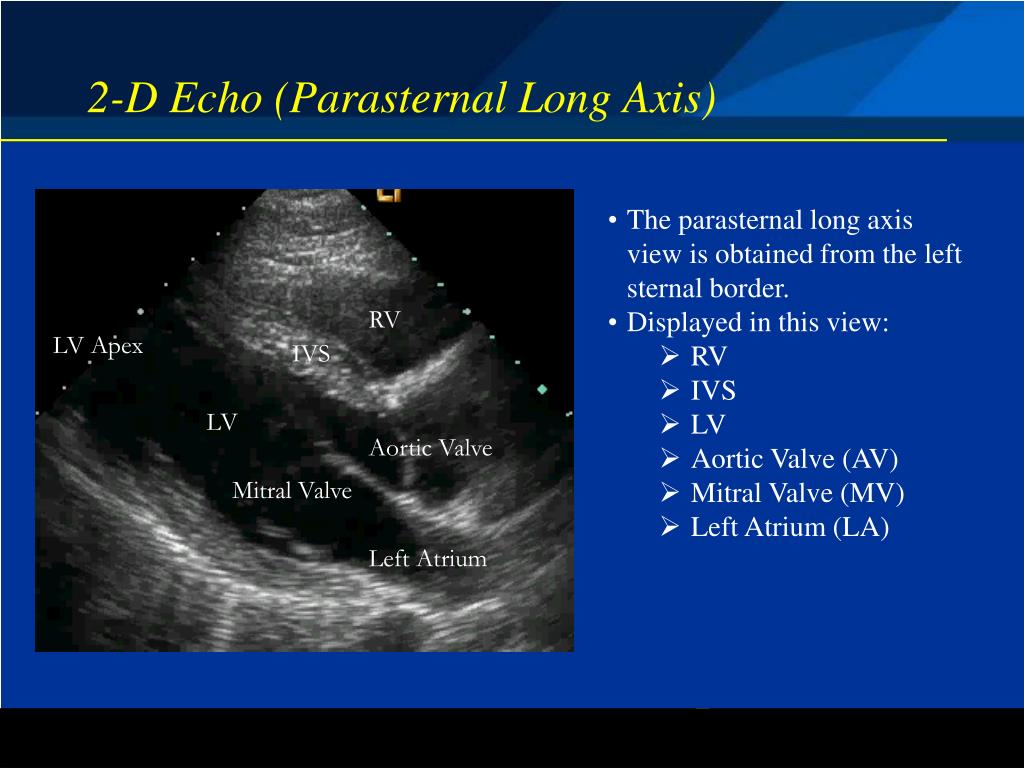
PPT - Basic Echocardiography Modes and Modalities PowerPoint Presentation -  ID:4979095
PPT - Basic Echocardiography Modes and Modalities PowerPoint Presentation - ID:4979095

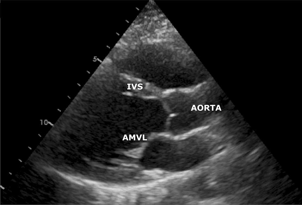
Interventricular septum (IVS) and left ventricle (LV) shapes in... |  Download Scientific Diagram
Interventricular septum (IVS) and left ventricle (LV) shapes in... | Download Scientific Diagram

Routine Echocardiogram Protocol With Standard 2D Echo Images and Color  Doppler - YouTube
Routine Echocardiogram Protocol With Standard 2D Echo Images and Color Doppler - YouTube

Progressive Thinning of the Basal Interventricular Septum by Giant Cell  Myocarditis - ScienceDirect
Progressive Thinning of the Basal Interventricular Septum by Giant Cell Myocarditis - ScienceDirect

Apical 4 chamber view - normal (transthoracic echocardiography) | Radiology  Case | Radiopaedia.org
Apical 4 chamber view - normal (transthoracic echocardiography) | Radiology Case | Radiopaedia.org

Number 18-04: Swollen inter-ventricular septum: A Phenotype mimicking HCM.  - Society for Cardiovascular Magnetic Resonance
Number 18-04: Swollen inter-ventricular septum: A Phenotype mimicking HCM. - Society for Cardiovascular Magnetic Resonance

Echocardiography - Wikiwand

Equine Echocardiography - Veterinary Clinics: Equine Practice

Echo M-mode With Pathologic Findings - Echocardiography | Echo, Pleural  effusion, Valvular
Echo M-mode With Pathologic Findings - Echocardiography | Echo, Pleural effusion, Valvular

Transthoracic echocardiography, bidimensionnal mode, standard... | Download  Scientific Diagram
Transthoracic echocardiography, bidimensionnal mode, standard... | Download Scientific Diagram

Echo pulmonary atresia with intact VS

Echocardiography | SpringerLink

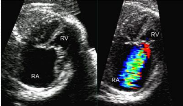
severe-tr-in-pa-ivs – SujyotHeartClinic

Short-axis 2-D ECHO (top) and corresponding M-mode ECHO (below) of the... |  Download Scientific Diagram
Short-axis 2-D ECHO (top) and corresponding M-mode ECHO (below) of the... | Download Scientific Diagram

A) Hypertrophic IVS and LVPW at the parasternal long-axis view by... |  Download Scientific Diagram
A) Hypertrophic IVS and LVPW at the parasternal long-axis view by... | Download Scientific Diagram

M-Mode Echocardiographic Measurements – All About Cardiovascular System and  Disorders
M-Mode Echocardiographic Measurements – All About Cardiovascular System and Disorders

A) Interventricular septal (IVS) imaging after 1 ml of myocardial... |  Download Scientific Diagram
A) Interventricular septal (IVS) imaging after 1 ml of myocardial... | Download Scientific Diagram

Right ventricular hypertrophy and hpertrabeculation misdiagnosed as a right  ventricular mass on echocardiography - ScienceDirect
Right ventricular hypertrophy and hpertrabeculation misdiagnosed as a right ventricular mass on echocardiography - ScienceDirect

View Image
View Image